Behzad Mokhtare1,
Meltem Cetin2 ![]() ,
Rukiye Sevinc Ozakar1,
Hatice Bayrakceken2
,
Rukiye Sevinc Ozakar1,
Hatice Bayrakceken2
For correspondence:- Meltem Cetin Email: melcetin@atauni.edu.tr Tel:+904422315236
Received: 6 August 2015 Accepted: 15 October 2016 Published: 26 February 2017
Citation: Mokhtare B, Cetin M, Ozakar RS, Bayrakceken H. In vitro and in vivo evaluation of alginate and alginate-chitosan beads containing metformin hydrochloride. Trop J Pharm Res 2017; 16(2):287-296 doi: 10.4314/tjpr.v16i2.5
© 2017 The authors.
This is an Open Access article that uses a funding model which does not charge readers or their institutions for access and distributed under the terms of the Creative Commons Attribution License (http://creativecommons.org/licenses/by/4.0) and the Budapest Open Access Initiative (http://www.budapestopenaccessinitiative.org/read), which permit unrestricted use, distribution, and reproduction in any medium, provided the original work is properly credited..
Purpose: To prepare metformin HCl-loaded alginate (AL) and alginate-chitosan (AL-CS) beads for oral application and to evaluate their in vitro characteristics and in vivo activities.
Methods: AL and AL-CS beads were prepared using ionotropic gelation. The beads were evaluated for particle size, surface morphology, drug encapsulation efficiency (EE) and in vitro drug release. The anti-diabetic effects of the beads were evaluated in diabetic Sprague Dawley rats.
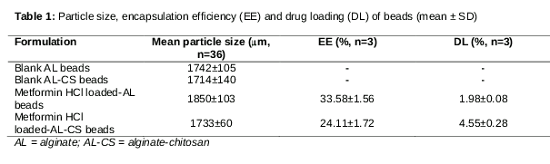
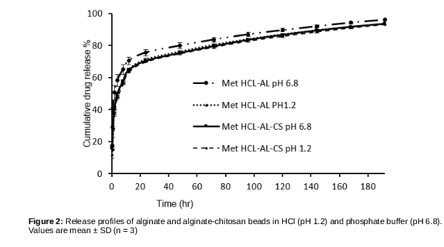
Results: The mean particle sizes of AL and AL-CS beads in wet state ranged from 1714 ± 140 to 1850 ± 103 µm. The EE % of AL and AL-CS beads were 33.58 ± 1.56 and 24.11 ± 1.72, respectively, with sustained in vitro drug release of about 93 to 96 % within 8 days in phosphate buffer (PB). Optimized metformin HCl-loaded AL and AL-CS beads showed significant hypoglycaemic effects in diabetic rats over a prolonged period (about 12 h) after oral administration compared to the pure drug (p < 0.05).
Conclusion: Metformin HCl-loaded AL and AL-CS beads for oral application may be useful in prolonging the hypoglycaemic effect of metformin. This is capable of increasing patients’ compliance to the medication.
Introduction
Metformin hydrochloride is a biguanide derivative widely used for the treatment of Type 2 diabetes, and prescribed for about 120 million people worldwide [1-4]. Metformin is recommended by the European Association for the Study of Diabetes, and the American Diabetes Association as the first line therapy for Type 2 diabetes [2,3]. Following oral administration, the drug is mainly absorbed from the upper small intestine, and has a relatively low bioavailability (absolute bioavailability of metformin is about 50 to 60 %). Its biological half-life (t1/2) is in the range of 0.9 – 2.6 h. Therefore, repeated applications of high doses of the medicine are needed for effective treatment. As a result, patient compliance is reduced and/or the incidence of side effects such as diarrhoea, nausea, anorexia, vomiting, weight loss, and taste disturbance are increased [1,5]. Moreover, lactic acidosis, which is sometimes fatal, has occurred with biguanides [1]. Its incomplete absorption is improved by using convenient drug delivery systems such as bio-adhesive and gastro-retentive drug delivery systems [6,7]. In addition, the development of different types of formulation for metformin is necessary for reduction of dosing frequency of the drug and its gastrointestinal side effects [8].
Sodium alginate, a salt of alginic acid, is a natural, polyanionic, non-toxic water-soluble copolymer of α-L-gluronic acid and β-D-mannuronic acid residues. It is obtained from marine brown algae and has the ability to form a gel network in the presence of divalent cations such as calcium in aqueous media. Thus, alginate beads are successfully prepared by ionotropic gelation [9,10]. More effective beads for the controlled release of drugs can be prepared using the combination of chitosan and alginate. The poly-electrolyte complex is formed with the ionic interaction between the amino residues of chitosan and the carboxyl residues of alginate [10]. The addition of chitosan in the formulation alters the diffusion rate of the encapsulated drugs and causes the bulk modification of the alginate bead structure [11]. CS is a polysaccharide obtained by the de-acetylation of chitin found in the exoskeleton of crustaceans (e.g. crab and shrimp) [12,13]. It has cationic character due to its primary amino groups and thus, shows muco-adhesive properties. AL beads and CS beads containing metformin HCl were prepared in previous studies [14,15]. In our study, besides metformin HCl-loaded AL beads, we also prepared AL-CS beads containing metformin HCl using AL and CS combination. CS and AL are very useful in pharmaceutical applications and they are the most widely used hydrogels in the preparation of sustained release dosage forms [11,15,16].
The present study focuses on the development and in vitro characterization of metformin HCl-loaded AL beads and AL-CS beads. It also focuses on in vivo evaluation of the hypoglycaemic activities of the bead formulations in streptozotocin (STZ) and nicotinamide-induced diabetic rats.
Methods
Materials
Metformin HCl was a generous gift from Sandoz Ilac Sanayi & Ticaret AS (Istanbul, Turkey). Sodium Alginate, STZ, nicotinamide and calcium chloride were obtained from Sigma-Aldrich (USA), and chitosan (Protosan UP CL 113) was purchased from FMC BioPolymer (Norway). All other chemicals and reagents were of analytical grade and used as they were received.
Preparation of metformin HCl-loaded AL and AL-CS beads
Metformin HCl-loaded AL and AL-CS beads were prepared by ionotropic gelation technique. Metformin HCl (100 mg) was dissolved in a sodium alginate aqueous solution (10 mL; 2 % w/v). AL-drug solution (bubble-free) was dropped through a 26-gauge syringe needle into 15 % (w/v) of calcium chloride solution on magnetic stirrer at 700 rpm for 15 min [17].The formed beads were further mixed in gelling medium at room temperature. After filtration, the beads were lyophilized for 24 h.
In addition, the AL-CS beads were also prepared by ionotropic gelation technique. Metformin HCl (100 mg) was dissolved in a sodium alginate aqueous solution (10 mL; 2 % w/v). The solution was dropped through a 26-gauge syringe needle into 5 % (w/v) of calcium chloride solution (pH adjusted to 4.5) containing chitosan (0.5 %, w/v) on magnetic stirrer at 700 rpm for 15 min. The resultant AL-CS beads were further mixed in gelling medium at room temperature [18,19]. After filtration, the beads were also lyophilized for 24 h.
Surface morphology and bead size
The surface morphology and shape evaluation of the bead formulations were examined by Scanning Electron Microscope (SEM; Inspect S50, FEI, USA) (used for lyophilized beads) and digital photographs (used for wet beads). The size of wet beads was determined by Vernier callipers.
Drug content of the beads
Lyophilized beads (20 mg) in 20 mL of PB (pH 6.8) in amber-coloured vials were sonicated for 15 min and mixed at 750 rpm for 4 hr in the dark for complete extraction of metformin HCl. The dispersion was then centrifuged at 5000 rpm for 15 min at 15 °C. The drug content of the supernatant of each sample was then measured using a validated UV method at 232 nm [20]. The experiment was performed in triplicate.
In vitro release studies
An incubation method was used for the investigation of metformin HCl release from beads in both media (PB, pH 6.8 and HCl, pH 1.2). Lyophilized beads (20 mg) were suspended in 20 mL of PB (pH 6.8) or HCl (pH 1.2) in amber-colored vials and then immersed in a constant temperature (37 °C) water bath. At pre-determined time intervals, samples (3 mL) were withdrawn from the release medium and replaced with the same volume of fresh buffer. All samples were centrifuged at 12500 rpm for 10 min, and their drug contents were measured using a validated UV method at 232 nm (for PB pH 6.8) and 209 nm (for HCl pH 1.2) [20]. The experiment was performed in triplicate.
FT-IR analysis
A Perkin-Elmer Spectrum One model FT-IR was used to record the IR spectra of AL, CS, metformin HCl, blank and metformin HCl-loaded beads prepared in KBr disks in the region of 4000–400 cm-1.
In vivo studies
The animal experiments were conducted according to the ethical norms approved by the Ethics Committee of Ataturk University (May 31 2013, No: 38). Fifty-nine male Sprague-Dawley rats (weighing 180 – 250 g) were obtained from the Experimental Animal Teaching and Research Center of the Experimental Animal Laboratory at Ataturk University. The rats were maintained on a normal diet and drinking water ad libitum at 24 ± 2 °C and under a 12 h light/dark cycle.
Analysis of fasting serum insulin levels
The basal blood glucose levels of overnight-fasted animals were determined using a glucometer (On.Call® Plus, Acon Lab, Inc, USA). Thereafter, diabetes was induced in the rats by administering a single intraperitoneal (i.p.) injection of freshly-prepared streptozotocin (STZ, 40 mg/kg body weight (b.w.)) in cold citrate buffer (pH 4.5). After 15 min, the rats were given freshly prepared nicotinamide solution i.p. (120 mg/kg b.w) [21]. Diabetes was confirmed in the rats by measuring blood glucose levels on day 7 of the induction. Animals with fasting blood glucose higher than 126 mg/dL were considered to be diabetic. The blood samples of diabetic rats (n = 8) collected from the tail veins were centrifuged (10,000 rpm, 10 min) and the supernatant was frozen until assayed for fasting serum insulin level using rat insulin enzyme immunoassay kit (SPI-BIO Bertin Pharma, France). The serum samples of control rats (healthy rats; n=8) were also analysed for fasting insulin level [21-23].
Evaluation of hypoglycaemic activity of beads in STZ-nicotinamide –induced diabetic rats
The rats were divided into six different groups as follows:
Group I: Control rats (healthy rats; n = 6)
Group II: Diabetic control (n = 8)
Group III: Diabetic rats treated with pure metformin HCl (100 mg/kg b.w) (n = 6)
Group IV: Diabetic rats treated with blank beads (n = 8)
Group V: Diabetic rats treated with metformin HCl-loaded AL-CS beads (equivalent to metformin HCl 100 mg/kg b.w.) (n = 9)
Group VI: Diabetic rats treated with metformin HCl-loaded AL beads (equivalent to metformin HCl 100 mg/kg b.w.) (n=6)
Type-2 diabetes was induced and controlled as described above. Diabetic rats were divided randomly into five different groups (Group II-Group VI).The pure drug and the formulations of blank and metformin HCl-loaded beads were administered orally through a flexible plastic tube after an overnight fast. Fasting blood samples were collected through the tail vein of the rats at onset, then on the 2nd, 3rd, 4th, 6th, 8th and 12th hr after the treatment, and analysed for glucose using a glucometer. Changes in body weight for all experimental rats were monitored. The rats remained fasted throughout the experiment, but were allowed free access to water.
Statistical analysis
The experimental data are expressed as mean ± SD. Statistical evaluations were performed using Mann-Whitney U test (SPSS Statistics 20.0 program; SPSS Inc, IL, USA). P < 0.05 was taken as indicative of statistical significance.
Results
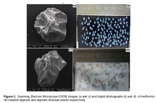
The SEM images and the digital photographs of metformin HCl-loaded AL and AL-CS beads (in dry and wet state, respectively) were shown in . They were spherical in the wet state (b and 1d). The values for particle size (in wet state) and EE % of all bead formulations are summarized in while the in vitro drug release profiles were given in . Regardless of the release medium, about 25 – 40 % of the loaded metformin HCL was released from the beads during the initial burst release (60 min). In addition, more than 93 % of the drug was released from both formulations in both media within 8 days ().
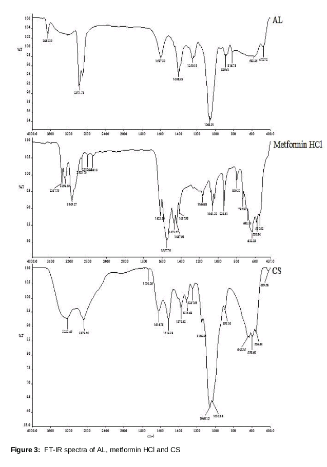
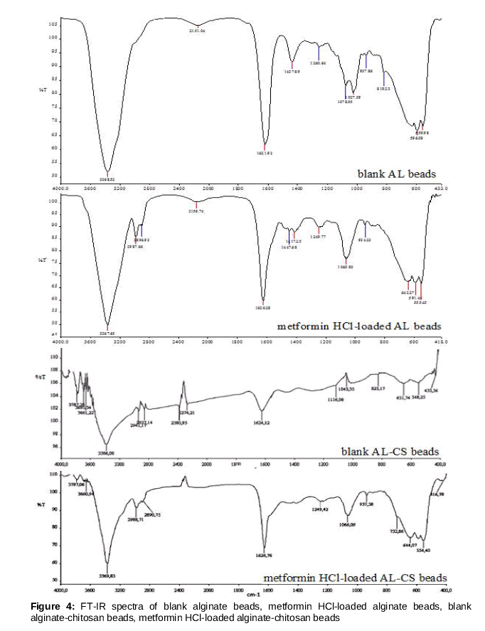
The FT-IR spectra of metformin HCl, AL, CS, and all bead formulations are presented in and . The FT-IR spectrum of AL polymer showed peaks at 3662.30 cm-1 due to O-H stretching, at 2971.71 cm-1 because of aliphatic C-H stretching, at 1406.38 cm-1 due to symmetric and asymmetric carboxylate salt groups stretching, at 1066.01 cm-1 related to C–O stretching, and at 889.51 cm-1 due to C-C and C-C-H stretching () [24]. The spectrum of metformin HCl displayed peaks at 3367.79 cm-1 (N-H asymmetric stretching), at 3293.10 cm-1 and 3149.27 cm-1 (N-H symmetric stretching), at 1621.90 cm-1 (C=N stretching), at 1557.70 cm-1 (N-H bending in plane), at 1472.57 cm-1, at 1447.01 cm-1 and 1417.93 cm-1 (C-H asymmetric bending; -CH3), at 1166.68 cm-1 and 1061.30 cm-1 (C-N stretching), at 936.41 cm-1 and 735.82 cm-1 (N-H wagging), at 632.29 cm-1 (NH2 rocking) and also at 550.02, 530.34 cm-1 (C-N-C bending) () [25,26]. Besides, the spectrum of CS presented peaks at 3232.49 cm-1 assigned to NH2 and O-H stretching, at 2879.55 cm-1 because of C-H stretching (CH2), at 1616.78 cm-1 attributed to C=O stretching, at 1146.85 cm-1 due to bridge-O stretching, at 1032.16 cm-1 and 1060.13 cm-1assigned to C-O stretching, and at 1373.42 cm-1 due to NHCO stretching () [27-29]. In the spectrum of metformin HCl-loaded AL beads, the bands in 2987.66 cm-1, 2896.93 cm-1, 1417.25 cm-1, 642.27 cm-1 related to the presence of metformin HCl were observed compared to the spectrum of blank AL beads (). Besides, the spectrum of AL-CS beads containing drug showed that the intensity of the peaks attributed to vibrations in the spectrum of drug (3369.83 cm-1, 2988.71 cm-1, 1626.76 cm-1, 1066.09 cm-1) increased and also bands were observed at 937.58 cm-1 and 732.86 cm-1 (). The FT-IR spectra of drug-loaded beads confirmed the presence of metformin HCl in both bead formulations.
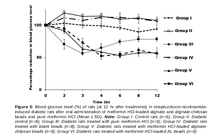
The fasting serum insulin levels of control rats and diabetic rats with fasting blood glucose level of higher than 126 mg/dL were found to be 0.66 ± 0.09 ng/mL and 0.51 ± 0.15 ng/mL, respectively. The reduction of fasting blood glucose level (as percentage of basal levels) after the administration of pure drug, and bead formulations versus time curves are given in . The fasting blood glucose concen-tration of Group II was higher than that of Group I (p < 0.05; ). Hypoglycaemic effect obtained for Groups V and VI were significantly different from that of Group III (p<0.05; except at 3 and 4 hr for Group V, and 4 hr for Group VI). However, there was no a significant difference between the hypoglycaemic effects of both bead formulations (p > 0.05).
Discussion
The results obtained in this study showed that the presence of chitosan in bead formulation had no significant effect on the size and drug content of the beads. Besides, the pH of the release medium had an effect on the drug release from AL beads, and a long-term hypoglycemic effect was observed in diabetic rats after oral administration of AL- and AL-CS- beads. The AL- and AL-CS-beads shrunk after freeze-drying, which is similar to the results reported by Zohar-Perez et al [30]. There was a slight but insignificant difference between particle sizes of the blank and drug-loaded AL and AL-CS beads, indicating that the presence of chitosan and incorporation of metformin HCl into the beads did not influence the particle size. This observation is in a good agreement with the results of Rajendran and Basu, who reported that size of AL-beads was not significantly enhanced by the addition of chitosan to the formulation [11]. The low EE% values obtained for the beads might be due to the high solubility of metformin HCl in water resulting in drug leakage to the aqueous medium. Similar observation was also made by Ramteke et al [31]. These authors reported low encapsulation efficiency (about 25 %) for metformin HCl-loaded alginate beads prepared using CaCl2, an observation attributed to the high aqueous solubility of drug [31].
All drug release profiles displayed a biphasic release pattern characterized by an initial fast release (burst effect), followed by a slower release rate. The initial burst release is attributable to the high water solubility of the drug and its adsorption to the surface of the beads. In addition, although drug release from the AL-CS beads were very similar in both release media (28 vs. 27 % for pH 6.8 and pH 1.2 in the first hour), drug release from the AL beads seemed to be influenced slightly by the pH of the medium. The pH of the release medium affects the swelling of AL beads and the drug release from AL beads. In an acidic release medium, the reduction in the electrostatic repulsion due to the formation of COOH groups and intermolecular hydrogen bond results in the restriction of the relaxation of polymer chains and formation of more compact network and thereby reducing drug release. It was observed that the degree of swelling of AL beads increased with increase of pH of release medium, probably due to exchange of the Ca2+ ion with Na+. These results are in agreement with the results of previous studies [11,15].
Various approaches (chemicals, dietary manipulation, or surgery) are available in the literature for development of experimentally-induced type 2 diabetes. For this purpose, we used STZ-nicotinamide. STZ taken up by pancreatic β-cells via GLUT2 (the glucose transporter) increases the production of free radicals and consequently causes impaired insulin secretion due to reduced insulin synthesis in β-cells and also the death of β-cells. On the other hand, nicotinamide prevents the STZ-induced cytotoxicity. Tahara et al [22] reported that co-administration of nicotinamide (100 mg/kg b.w.) and streptozotocin (50 mg/kg b.w.) was successful in inducing mild diabetes in rats. Reduction in the fasting blood glucose level by the pure drug was obtained for 3 hr and the level restored after 3 hr. On the other hand, the fasting blood glucose levels of metformin HCl-loaded AL-CS and AL beads gradually declined and peaked in 6 hr and 4 hr, respectively, indicating that hypoglycaemic effect was extended by both bead formulations. Similar observations are reported in the literature [15,32]. Nayak et al [15] evaluated the hypoglycaemic effects of non-floating and floating alginate beads containing metformin HCl on STZ-induced diabetic rats and found that a significant hypoglycaemic effect was obtained for floating beads 3 to 9 hr after bead administration. However, this effect was visible up to 5 hr after the administration of non-floating beads. Thus, floating beads were more useful for sustained hypoglycaemic effect, and a 25 % reduction in the blood glucose level was considered as significant hypoglycaemic effect. Similarly, following administration of metformin HCl-loaded fenugreek seed mucilage alginate muco-adhesive beads (equivalent to 100 mg/kg metformin HCl) to alloxan-induced diabetic rats, a significant hypoglycaemic effect was demonstrated up to 10 hr, and about 30 % reduction in blood glucose level was obtained during this period [32]. These results support our observation with regard to the prolonged hypoglycaemic effect obtained with metformin HCl-loaded AL and AL-CS beads.
Conclusion
This study shows that HCl-loaded AL and AL-CS bead formulations may be useful in prolonging the hypoglycaemic effect of orally administered metformin. This is capable of increasing patient compliance with the medication. Further are, however, needed to explore the degree of side effect reduction of the formulated metformin.
Declarations
Acknowledgement
References
Archives


News Updates